Arthroscopic OCD Fixation

Arthroscopic OCD fixation stabilizes lesions, reduces pain, preserves joint function, and promotes cartilage healing with minimally invasive surgery.

99.8%

Safety Rate

0 +
Years of Experience
0 +
Dedicated Service
0 +
Surgeries
0 +
Happy Patients

Arthroscopic OCD Fixation Services

Comprehensive Arthroscopic OCD Fixation care with advanced monitoring and safety protocols

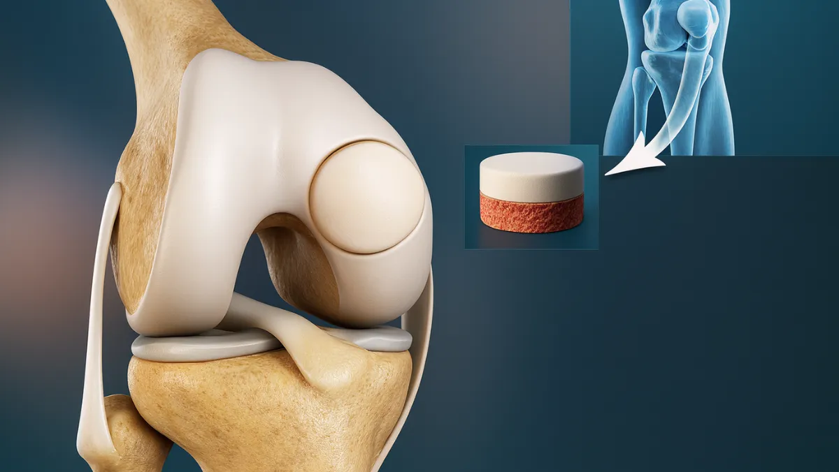
Osteochondritis Dissecans (OCD) is a joint condition where a segment of cartilage, along with a thin layer of underlying bone, becomes loose due to reduced blood flow. This commonly affects the knee joint, particularly the femoral condyle, and can cause pain, swelling, locking, and instability. If untreated, OCD may progress to long-term cartilage damage and early arthritis.

Arthroscopic OCD fixation is a minimally invasive surgical technique that stabilizes the loose fragment and restores the smooth joint surface. Unlike open surgery, arthroscopy uses small incisions and specialized instruments, allowing faster recovery, less scarring, and improved outcomes.

Why Arthroscopic OCD Fixation Is Performed

Surgery is recommended when:

  • The OCD lesion is unstable or partially detached.
  • Non-surgical treatments (rest, physiotherapy, activity modification) fail.
  • The patient is young and active, with a repairable lesion.
  • Symptoms such as pain, swelling, or knee locking persist.

Arthroscopic Surgical Procedure

  1. Diagnosis and Assessment
    MRI and X-rays confirm the size, location, and stability of the OCD lesion.
  2. Arthroscopic Access
    Small keyhole incisions allow insertion of a camera (arthroscope) and instruments into the knee.
  3. Preparation of the Lesion Site
    The unstable cartilage/bone fragment is identified, and the defect bed is cleaned to promote healing.
  4. Fixation of the OCD Fragment
    The loose fragment is repositioned and stabilized using bioabsorbable pins or screws, that hold it in place while healing occurs.
  5. Final Check and Closure
    The surgeon ensures the fragment is stable, joint motion is smooth, and then closes the tiny incisions with sutures or strips.

Recovery and Rehabilitation

Healing after arthroscopic OCD fixation requires a structured plan:

  • Immobilization & Protection: A brace and crutches are used initially to limit stress on the repair.
  • Gradual Weight-Bearing: Patients progress from partial to full weight-bearing as healing advances.
  • Physiotherapy: Exercises restore motion, strengthen surrounding muscles, and improve stability.
  • Return to Sports: Low-impact activities resume around 3–4 months, while full sports participation may take 6–9 months.

Benefits of Arthroscopic OCD Fixation

  • Preserves the patient’s natural cartilage and bone.
  • Minimally invasive, with smaller incisions and faster recovery.
  • Restores knee stability and reduces pain.
  • Lowers the risk of future arthritis compared to fragment removal.


Risks and Considerations

Though generally safe, potential risks include incomplete healing, hardware irritation, infection, or recurrent instability if the lesion fails to unite. Success rates are highest when performed early and followed by strict rehabilitation.

Summary

Arthroscopic OCD fixation is an effective, minimally invasive way to treat unstable cartilage lesions in the knee. By preserving and stabilizing the natural joint surface, it relieves pain, restores function, and protects long-term knee health—without the need for open surgery.


Frequently Asked Questions

Common questions about Arthroscopic OCD Fixation and our services

Related Blogs

No data was found

Don't Let Joint Pain Limit Your Life

Take the first step towards pain-free living. Book your consultation today and discover personalized treatment options tailored to your needs.

Book an Appointment